
|
2/11/2018 0 Comments Abdominal Wall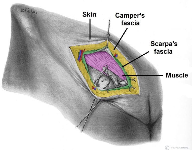 The abdominal wall encloses the abdominal cavity, and can be divided into anterolateral and posterior sections. Its key functions include:
Superficial FasciaThe superficial fascia consists of fatty connective tissue. The composition of this layer depends on its location:
Fig 1 – The layers of the anterolateral abdominal wall. Below the umbilicus, there are two layers of superficial fascia – Camper’s and Scarpa’s. Muscles of the Abdominal WallThe muscles of the anterolateral abdominal wall can be divided into two main groups:
In the anteromedial aspect of the abdominal wall, each flat muscle forms an aponeurosis(a broad, flat tendon), which covers the vertical rectus abdominis muscle. The aponeuroses of all the flat muscles become entwined in the midline, forming the linea alba (a fibrous structure that extends from the xiphoid process of the sternum to the pubic symphysis). External Oblique The external oblique is the largest and most superficial flat muscle in the abdominal wall. Its fibres run inferomedially.
The internal oblique lies deep to the external oblique. It is smaller and thinner in structure, with its fibres running superomedially (perpendicular to the fibres of the external oblique).
0 Comments
Leave a Reply. |
Search by typing & pressing enter
